

EngramBio
Repair, restore, remember
Repairing the synapses that hold memory together.
We develop therapeutics that repair the synaptic connections lost in Alzheimer's disease and related dementias, built on foundational neuroscience showing how the damaged brain can regain its ability to form new memories.
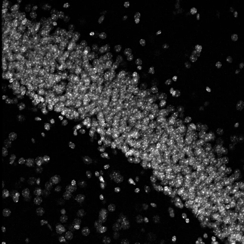
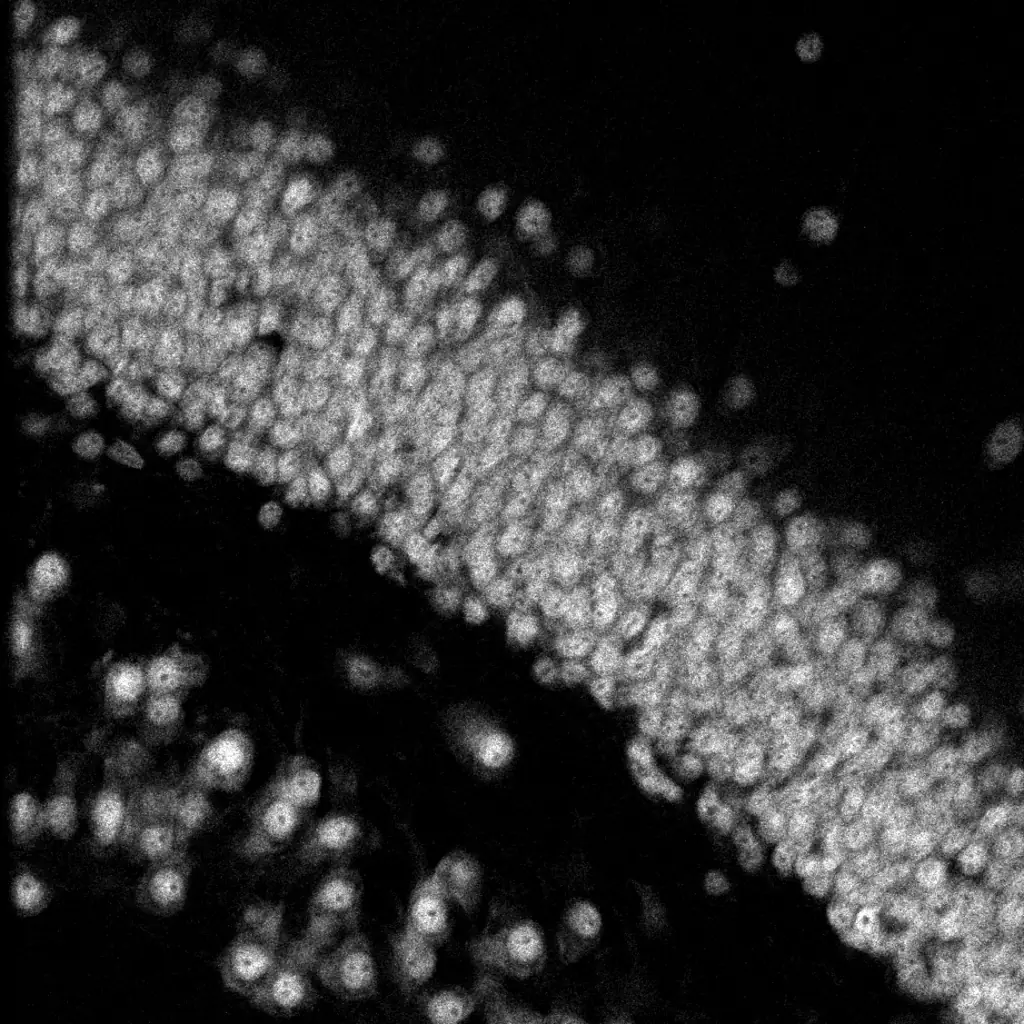
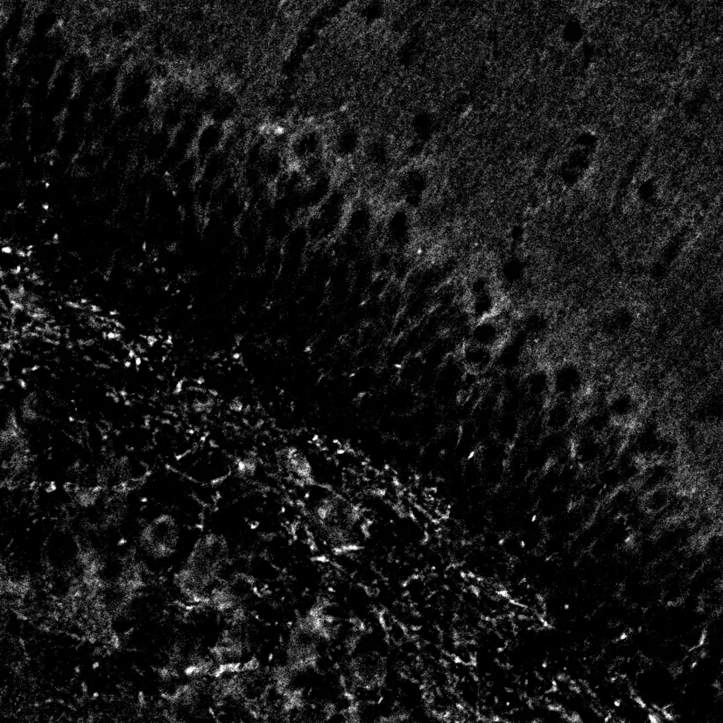
Fig. 1 · Example image of the mouse hippocampus (dentate gyrus), imaged with confocal microscopy. Cell nuclei (DAPI) in blue, neurons in coral, and neuron nuclei, where the two overlap, in pink; dendrites in gold.
ABOUT
A new class of therapeutics built to repair the synapse.
EngramBio engineers therapeutics that restore the synaptic connections lost to Alzheimer's disease, related dementias, and aging. Synapse loss, more than plaques or tangles, is the strongest correlate of cognitive decline in Alzheimer's disease. Our lead program builds on more than a decade of original neuroscience showing that a peptide derived from KIBRA repairs damaged synapses and reverses memory deficits in preclinical models.
Hippocampus · Fig. 1
Where memories form.
This is the mouse hippocampus, a brain region essential for forming new memories.
Hippocampus · Fig. 1 · Confocal
Hippocampus · Fig. 1 · Confocal
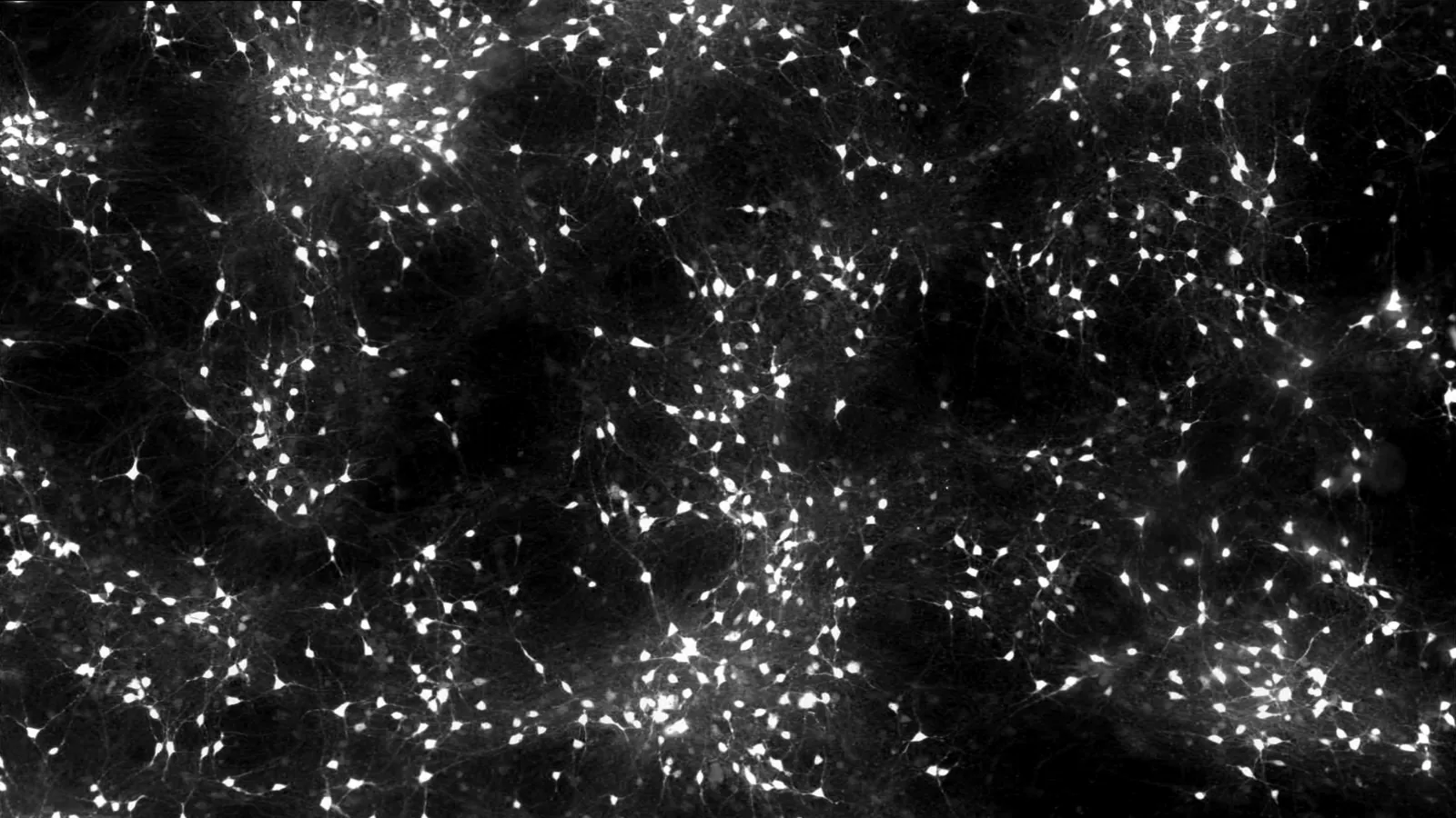
Fig. 2 · Example image of human iPSC-derived neurons in culture, imaged with fluorescence microscopy.
Network · Fig. 2
Neurons, connected.
Every memory depends on neurons reaching out and connecting to one another. Imaging lets us see those connections directly, including where they hold strong and where they start to break down in diseases like Alzheimer's. It's how we know whether a therapy is protecting the networks that matter most.
Seeing is understanding.
Much of what we know about the brain started with someone looking closely. Imaging lets us watch neurons grow and reach toward one another. The bright points are cell bodies, and the faint threads are the branches that connect them. Where those branches meet, they form synapses, the connections that learning and memory depend on.
Network · Fig. 2 · Widefield fluorescence
Network · Fig. 2 · Widefield fluorescence
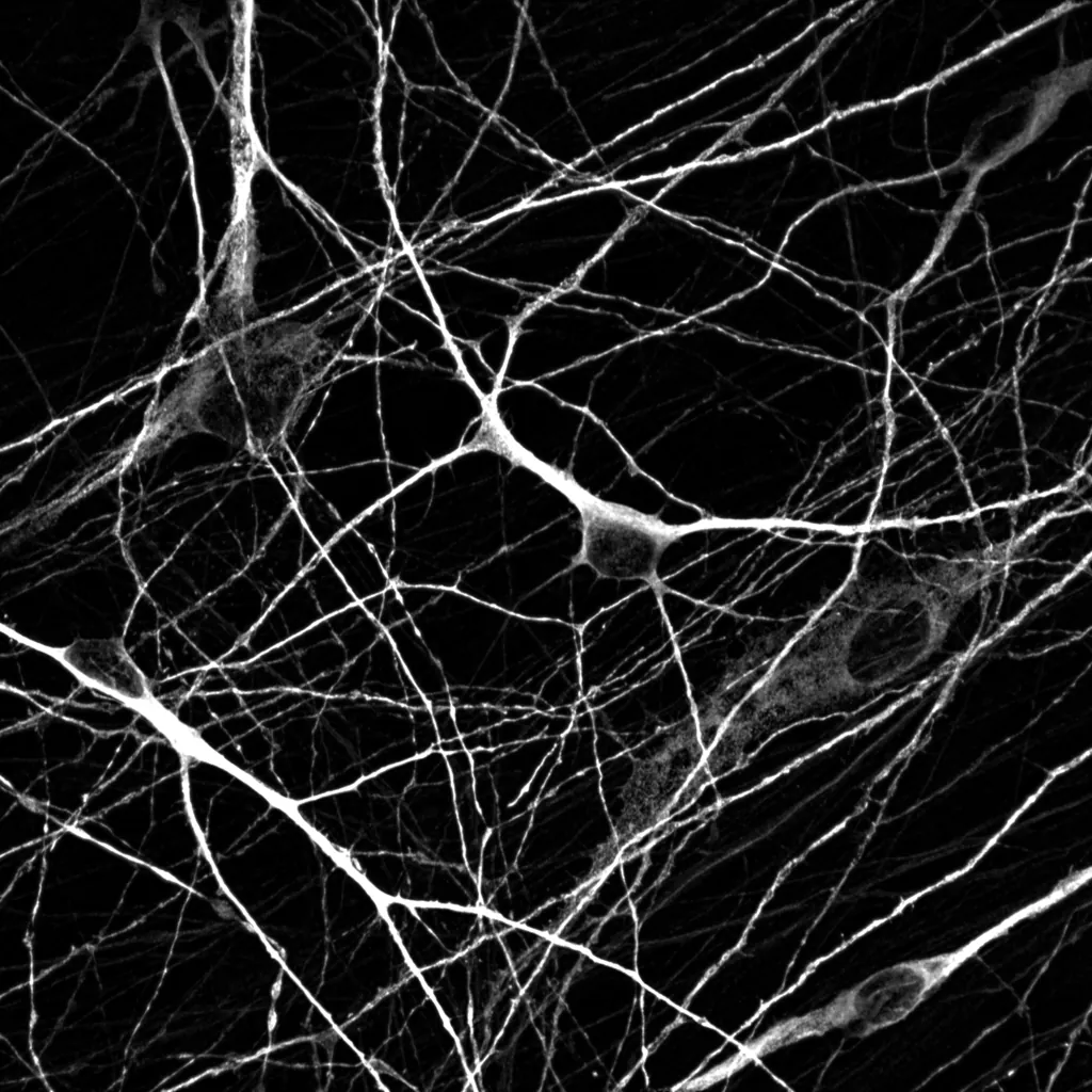
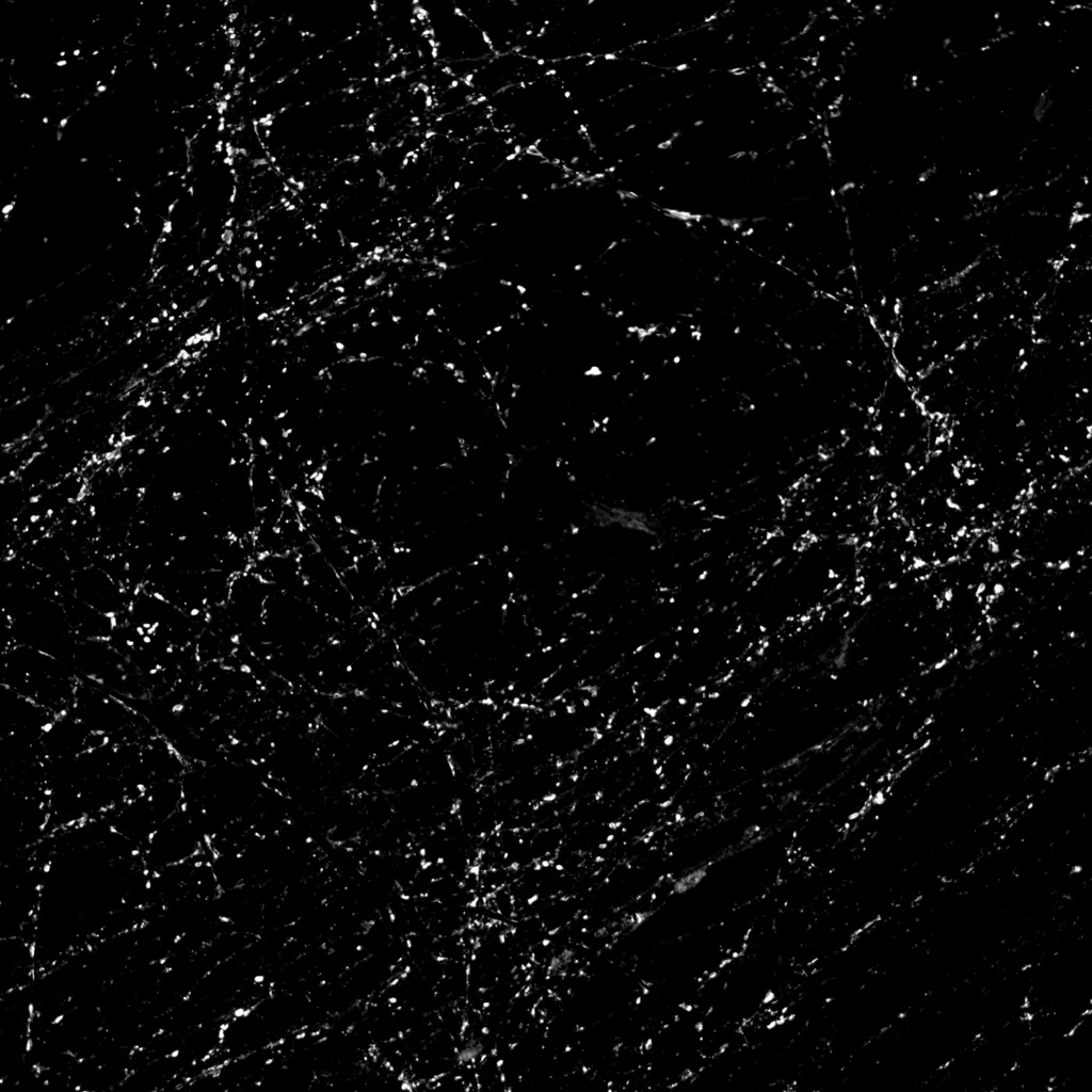
Fig. 3 · Neurons in culture, imaged with confocal microscopy. Dendrites in blue, synapses in gold.
Neuron · Fig. 3
Where neurons listen.
Closer in, we can follow the branches themselves. These are dendrites, the part of a neuron that receives signals from other neurons. Here, the dendrites are stained for MAP2, a protein that supports their internal skeleton, so only the dendrites and cell bodies are labeled. Along these branches, incoming connections form synapses, the sites where memory takes shape.
Neuron · Fig. 3 · Confocal
Where synapses sit.
A second stain marks synapsin, a protein of the nerve terminals where one neuron passes its signals to the next.
Neuron · Fig. 3 · Confocal
Neuron · Fig. 3 · Confocal
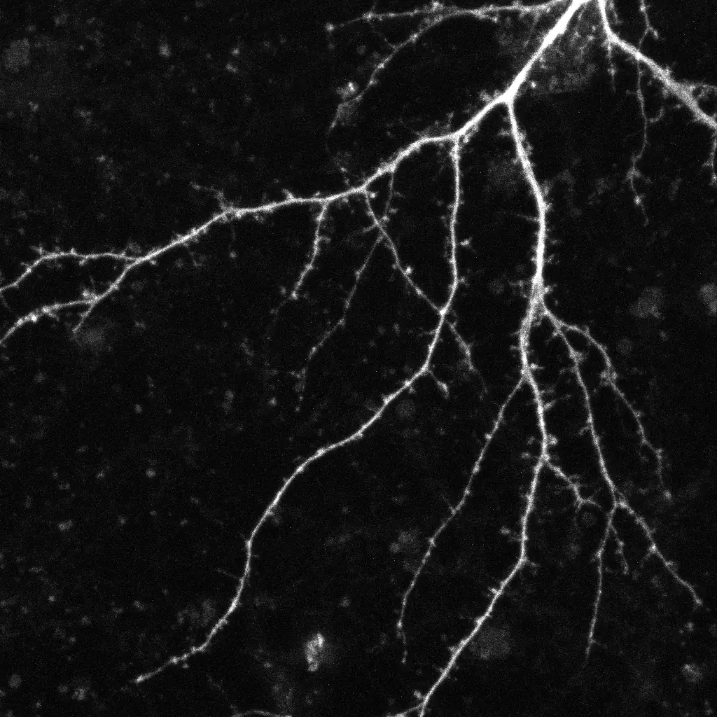
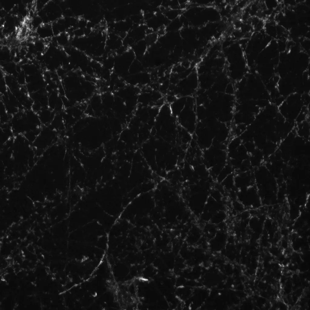
Fig. 4 · Rat hippocampal neurons in culture, imaged with confocal microscopy before and after long-term potentiation (LTP), a laboratory model of learning. After LTP, staining for actin, a structural protein that shapes dendritic spines, appears more intense. Dendrites in blue, actin in gold. Before and after are separate samples.
Dendrite · Fig. 4
Structure that can change.
Connections strengthen and weaken with experience, and dendritic spines, the small protrusions where most synapses sit, change shape with them. This capacity, called synaptic plasticity, underlies learning and memory. In Alzheimer's disease, pathogenic tau obstructs it, a key mechanism behind memory loss.
Dendrite · Fig. 4 · Confocal
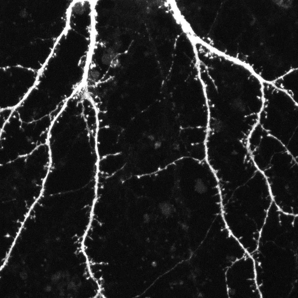
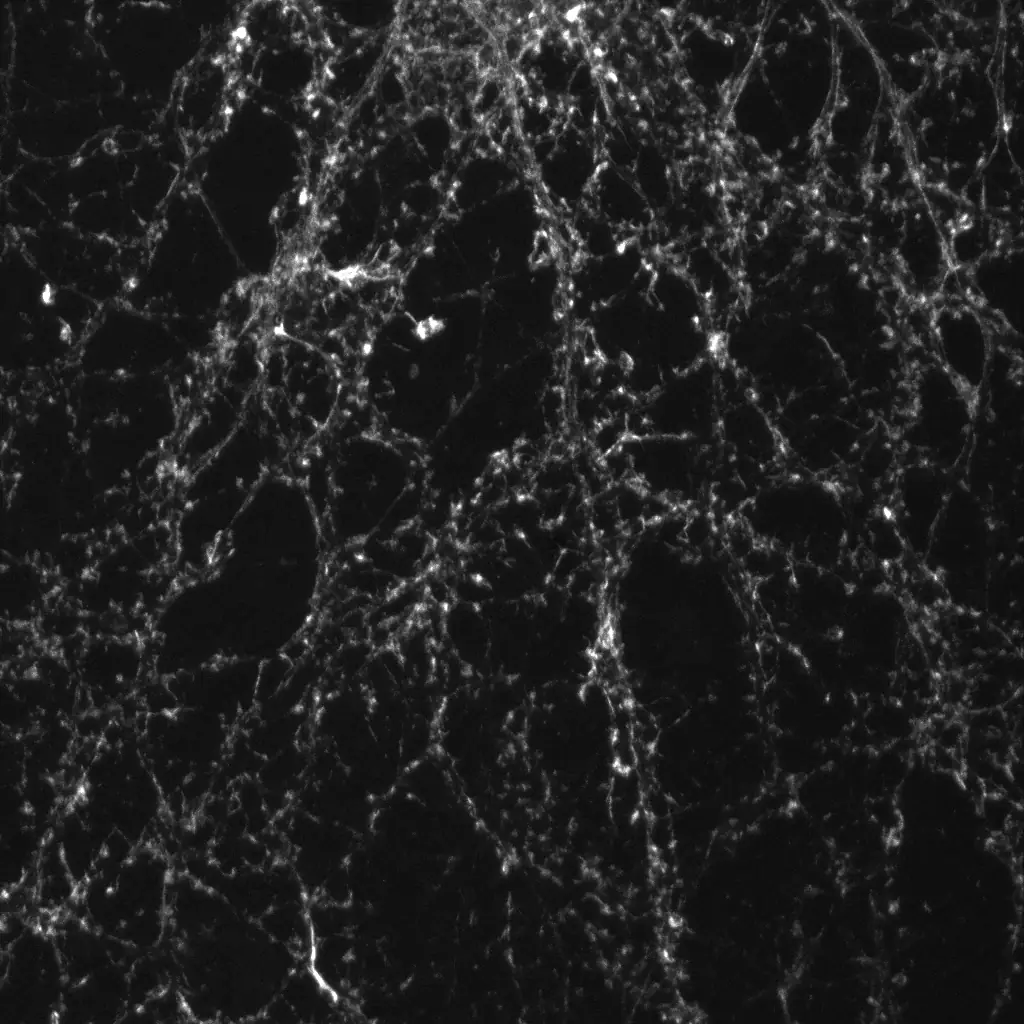
A trace of learning.
These images show neurons before and after long-term potentiation, a cellular model of learning and memory formation. Potentiation strengthens synapses, and the synapses themselves can grow larger.
Dendrite · Fig. 4 · Confocal
Dendrite · Fig. 4 · Confocal
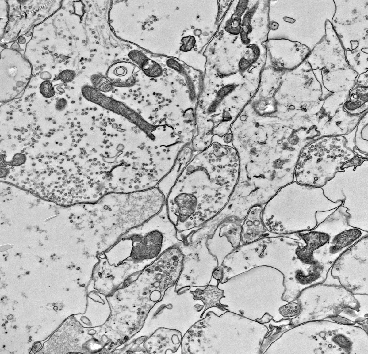
Fig. 5 · Neurons in culture, imaged with electron microscopy at 20,000× and 50,000×.
Synapse · Fig. 5
A connection you can see.
An electron microscope reveals detail far beyond the reach of light. At 20,000 times magnification, cell membranes appear as fine lines, and nerve terminals are packed with small round vesicles that carry chemical signals to the next neuron.
Synapse · Fig. 5 · Electron microscopy
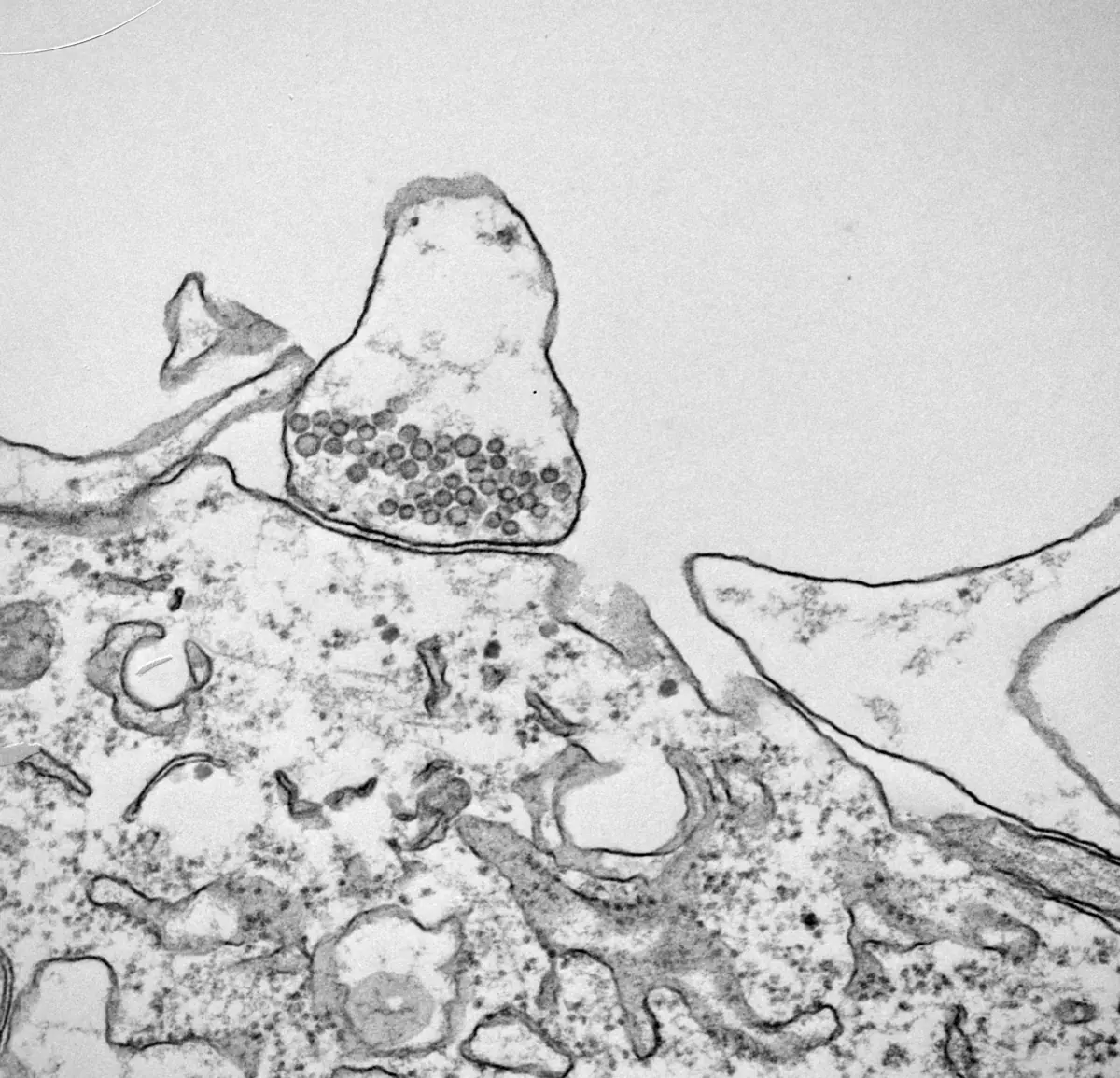
Vesicles carry the message.
At 50,000 times magnification, the vesicles appear as rings about 40 nanometers across. Each one holds neurotransmitters. When a signal arrives, vesicles at the release site fuse with the cell membrane, and their neurotransmitters cross a narrow gap, the synapse, to reach the next cell.
Synapse · Fig. 5 · Electron microscopy
Where the decline begins.
Alzheimer's doesn't begin with forgetting a name. It begins at the synapse. In human iPSC-derived neurons, a 30-minute exposure to tau oligomers set off a progressive decline on both sides of the synapse. Some synapses were lost. Others remained but stayed weakened, with fewer vesicle clusters at their release sites. Our founders co-authored this 2026 study.
Read the study (opens in a new tab)Synapse · Fig. 5 · Electron microscopy
Fig. 6 · Illustrative model of the proteins and receptors in one synapse, after the 2024 JCI paper. PKMζ in gold, KIBRA in teal, AMPA receptors in blue, pathogenic tau in coral; CT-KIBRA, a fragment of KIBRA, is outlined.
Molecule · Fig. 6
Learning adds receptors.
Firing at a synapse increases PKMζ, a protein in the receiving spine. PKMζ increases the AMPA receptors at the spine's surface, which catch the signal. More receptors make a stronger synapse, a sign of learning.
Molecule · Fig. 6 · Mechanism Illustrative
KIBRA keeps learning active.
PKMζ has a natural turnover: it is broken down and replaced over time. KIBRA binds to PKMζ and slows that turnover, so the receptors, and the learning they hold, stay in place.
Molecule · Fig. 6 · Mechanism Illustrative
Aging lowers KIBRA.
As we age, KIBRA decreases. In normal aging, that is fine: the synapse keeps its receptors.
Molecule · Fig. 6 · Mechanism Illustrative
Tau reduces receptors.
Pathogenic tau is different. It reduces AMPA receptors at the synapse's surface, and that leads to memory loss.
Molecule · Fig. 6 · Mechanism Illustrative
A fragment that acts like KIBRA.
CT-KIBRA, a fragment of KIBRA, acts the same way as KIBRA. It helps stabilize PKMζ, and the AMPA receptors lost to pathogenic tau are replaced. Tau itself stays.
Molecule · Fig. 6 · Mechanism Illustrative
Repair starts with biology.
KIBRA is a synaptic protein essential for learning and memory. It helps synapses strengthen during learning, the same plasticity shown in the images above. A genome-wide study of human memory first pointed to KIBRA, finding that common variants of the gene are linked to memory performance, and some later studies tied those variants to Alzheimer's disease risk. In mice expressing pathogenic human tau, a fragment of KIBRA restored synaptic plasticity and memory without changing tau levels. That 2024 finding, co-authored by our founders, is the foundation of our lead program and of our approach to synaptic resilience: helping synapses keep working, even in the presence of disease.
Read the paper (opens in a new tab)Molecule · Fig. 6 · Mechanism Illustrative
Molecule · Fig. 6 · Mechanism Illustrative
Fig. 7 · Guided design search · 12 / 12 committed. Illustrative model - computed for visualisation, not an experimentally validated design. On narrow screens, cropped to its central 4:3.
TECHNOLOGY
An integrated discovery engine for synapse-repair therapeutics.
Three disciplines, one decision at a time. Chemistry-aware modeling narrows a vast peptide design space. Physics-based simulation ranks candidates by binding fidelity and brain stability. Human iPSC-derived neuron assays then test every claim before a molecule moves forward.
Molecule · Fig. 7 · Computational model Illustrative
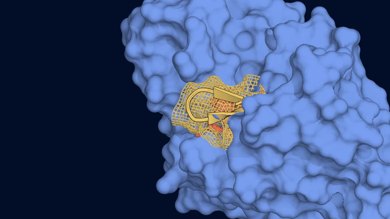
Chemistry-Aware Peptide Design
We design lead molecules using structure-prediction tools that natively model drug-like properties. Candidates are then refined under physics-based molecular dynamics and free-energy calculations and ranked across binding, brain stability, and clinical translatability simultaneously. The result is a pipeline that converges on molecules with the trade-offs already engineered in.
Neuroscience-Led Wet Lab Validation
Every lead candidate is validated by our neuroscience team in disease-relevant models such as human iPSC-derived neurons, using methods such as electrophysiology and multi-electrode array recording to assess synaptic plasticity and network connectivity, with follow-up behavioral studies. Every computational claim is checked against experimental measurements before a molecule advances.
Discovery Platform
We are turning the science behind our CT-KIBRA lead into a repeatable discovery process. It starts from human genetic data, public knowledge of synapse biology and our own data from patient-derived cells. Computational design proposes candidates, and laboratory screens and tests in neurons and in mice decide which advance. Our lead peptide is in hit-to-lead development, where promising molecules are tested and refined, and the platform is built to bring more candidates there.
See the pipelineMolecule · Fig. 7 · Computational model Illustrative
Repair the synapse. Restore the circuit. Remember.
Alzheimer's disease reaches past the synapse, into families, caregivers and communities. Every synapse we repair is a connection preserved.
AWARDS & PARTNERSHIPS

NIH NIA Startup Challenge Finalist
Selected as a finalist among top innovators nationwide for developing breakthrough technologies to advance healthy aging and address critical challenges in aging research.

NIH NIA Startup Challenge Winner
Awarded winner by the National Institute on Aging for our innovative approach to improving healthy aging outcomes, recognizing our technology's potential to transform aging care and research.

NVIDIA Inception
NVIDIA Inception is a program for early-stage startups that are using AI to solve real-world problems.
MEET THE TEAM
Leadership Team


Ready to Transform Drug Discovery?
Join us in accelerating the development of life-changing neurological therapeutics.
Whether you're a pharmaceutical partner, investor, or research collaborator, let's discuss how we can work together to bring hope to patients worldwide.
Get in touchAbout these images
The figures are example images. Fluorescence microscopes record each stain as brightness, so each is shown in a color assigned to it; Fig. 3's two stains were separated by color from one merged image, and its synapses are drawn over the dendrites rather than added to them. Electron micrographs are shown as recorded, in grayscale. Brightness and contrast are adjusted evenly across each whole image, and some figures are cropped to fit the screen. Scrolling reveals each image a stain or a field at a time, and the last frame is the image itself. Nothing is added to or removed from any image.